DeepLesionBrain
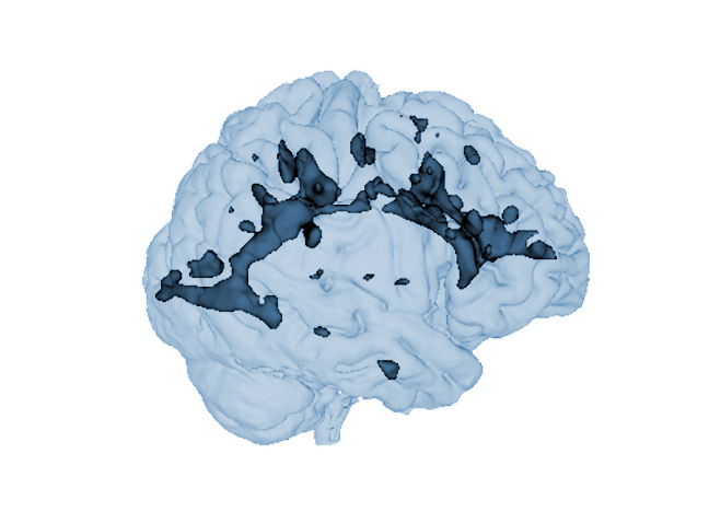
DeepLesionBrain is a pipeline to automatically segment white matter lesions from MRI data (T1w + FLAIR). It gets anonymized MRI brain volumes in NIFTI format and produces a pdf report with the volumes of the lesions and their locations. Moreover, DeepLesionBrain offers brain parcellation and disconnectome analysis.
Labelling protocol
White matter lesions were manually outlined by an expert radiologist using multimodal MRI data (T1w + FLAIR) to create a library of 43 cases. After the segmentation process, lesions are classified based on their location as periventricular, deep white, juxtacortical and infratentorial. After lesion inpainting, the T1w MRI is segmented using AssemblyNet. Finally, the detected lesions are used to estimate a disconnectome map based on the HCP1065 atlas.
Report
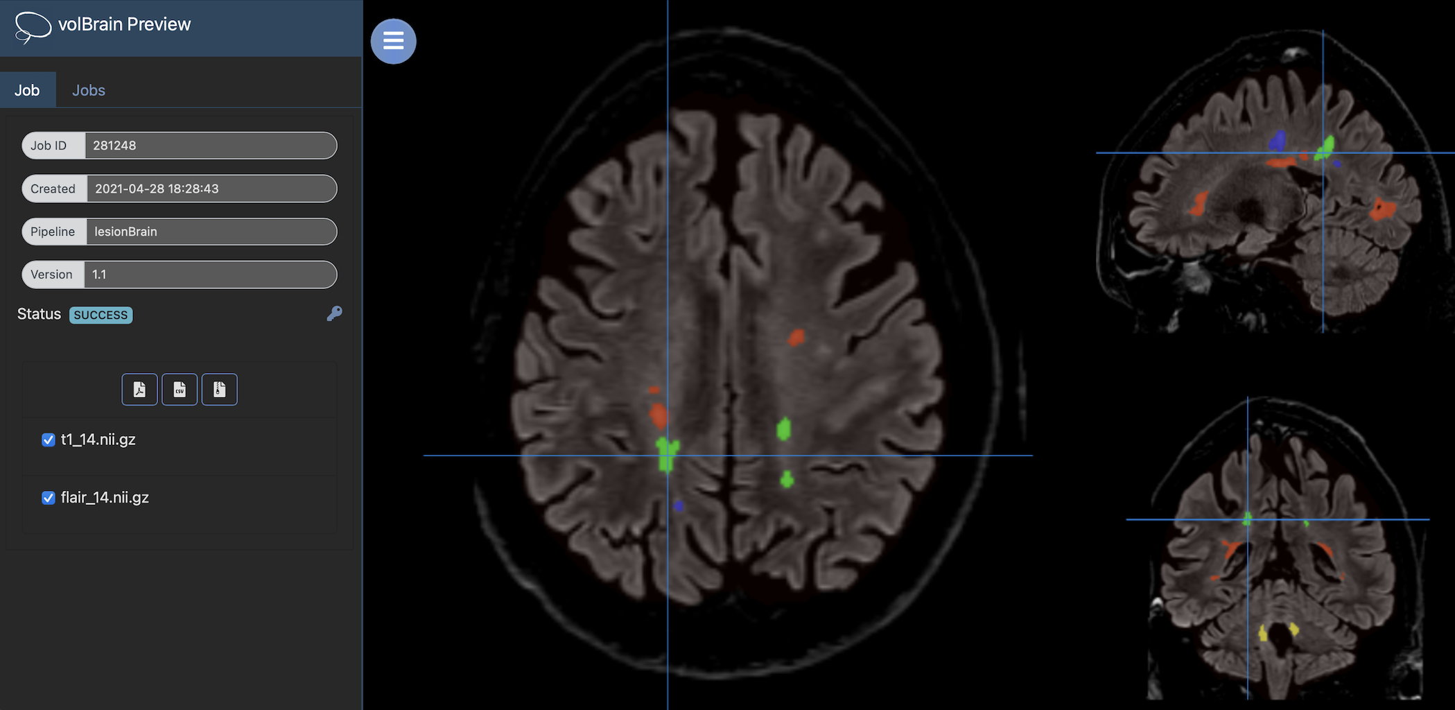
Once the process is finished, you will be notified by e-mail so you will be able to download a package including some image files and two reports (CSV and PDF) offering all the volumes estimated from the segmentations. As you can see in the figure below, the PDF includes patient information, lesion types, their volumes and locations in the MNI space. Moreover, the volumes of 132 brain structures based on AssemblyNet segmentation are provided. Finally, DeepLesionBrain provides the probability of disconnection caused by the detected lesions for 64 white matter tracts. It also includes several snapshots from the different labeling steps as a quality control.
Download PDF Report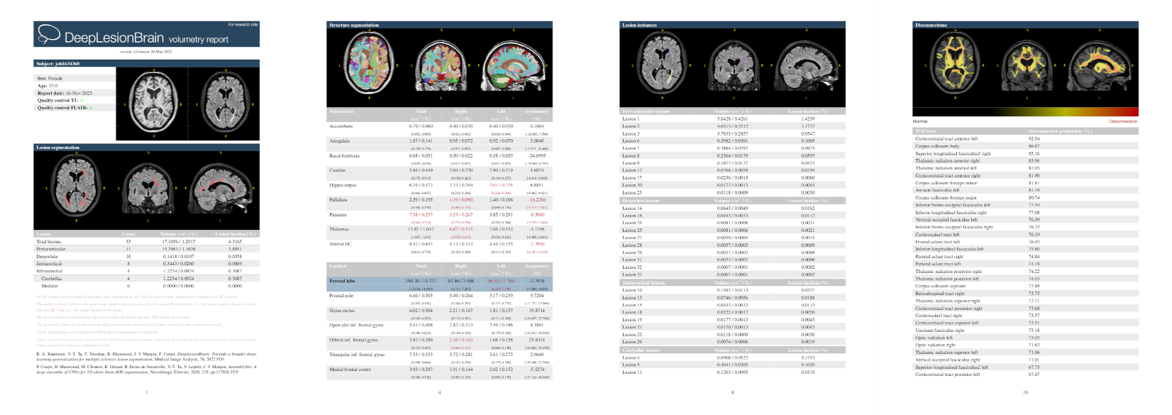
References
R. A. Kamraoui, V.-T. Ta, T. Tourdias, B. Mansencal, J. V. Manjón, P. Coupé. DeepLesionBrain: Towards a broader deep-learning generalization for multiple sclerosis lesion segmentation. Medical Image Analysis 76 (2022): 102312 PDF
P. Coupé, B. Mansencal, M. Clément, R. Giraud, B. Denis de Senneville, V.-T Ta, V. Lepetit, J. V. Manjón. AssemblyNet: A large ensemble of CNNs for 3D Whole Brain MRI Segmentation. NeuroImage, 219, 117026, 2020. PDF
J. V. Manjón, J. E. Romero, R. Vivo-Hernando, G. Rubio, F. Aparici, M. de La Iglesia-Vaya, T. Tourdias, P. Coupé. Blind MRI brain lesion inpainting using deep learning. SASHIMI workshop MICCAI 2020 PDF
de Senneville, B.D., Manjón, J.V. and Coupé, P., 2020. RegQCNET: Deep quality control for image-to-template brain MRI affine registration. Physics in Medicine & Biology, 65(22), p.225022. PDF