petBrain

petBrain is a pipeline of processes aimed to automatically analyze MRI and PET brain data. It gets an anonymized MRI and PET brain volumes in NIFTI format and produces a pdf report with A/T/N patient's status.
A/T/N model
petBrain is a novel pipeline designed for the fast, reliable, and standardized analysis of Alzheimer’s disease biomarkers. It enables end-to-end processing of amyloid-PET, tau-PET, and structural MRI using advanced deep learning algorithms. The pipeline simultaneously estimates amyloid (A), tau (T2), and neurodegeneration (N) biomarkers through established quantification frameworks such as Centiloid, Centaur, and HAVAs. As a result, petBrain provides outputs directly aligned with clinical and research standards.
Designed for accessibility, no local installation or technical expertise is required—users simply upload their data and receive high-quality results. petBrain achieves performance comparable to leading pipelines, showing strong agreement with fluid biomarkers, clinical status, and cognitive outcomes. This makes it a powerful tool for clinical research, biomarker-based patient stratification, and personalized medicine in Alzheimer’s disease.
Online Preview
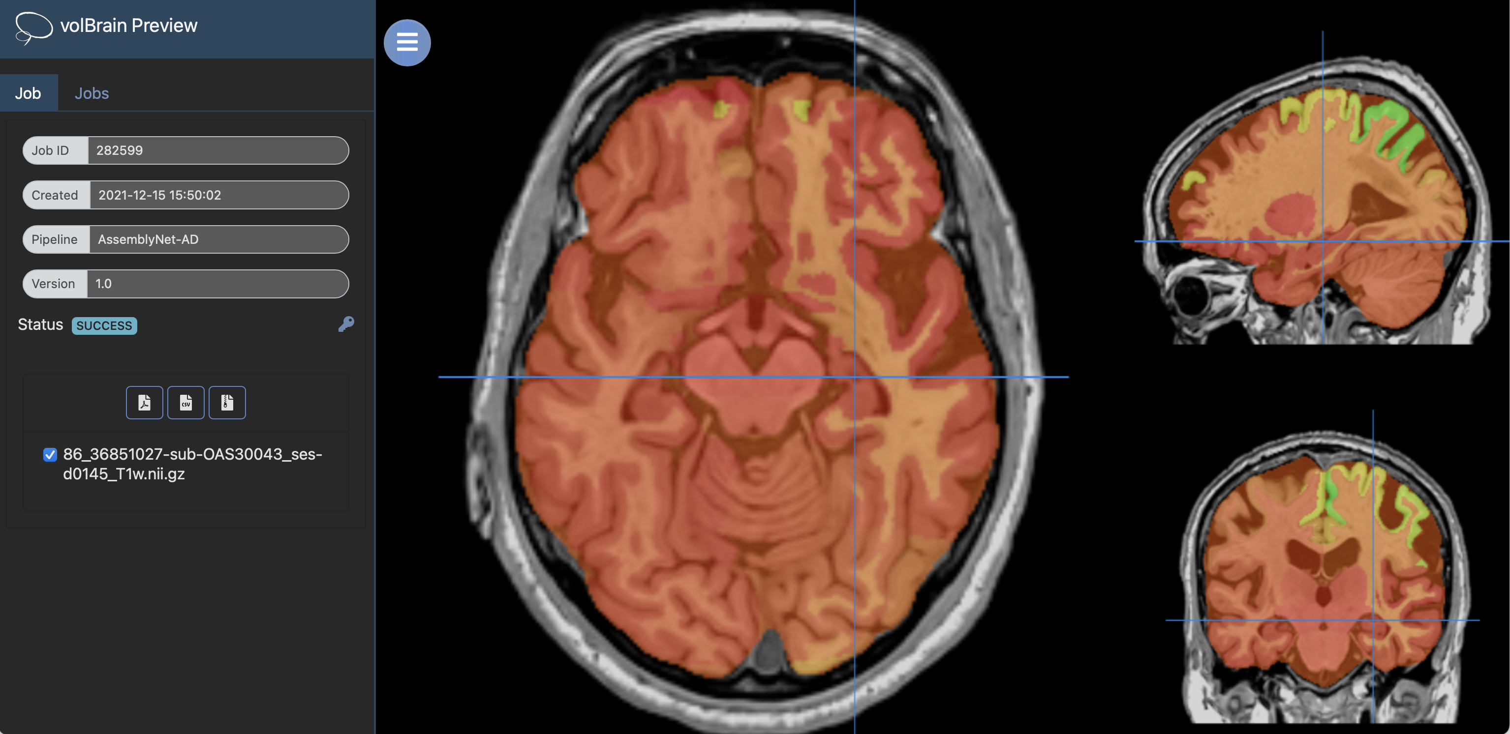
In the online preview , when both tau and amyloid PET are provided, tau-PET is overlaid on the T1-weighted image. When only one PET is uploaded, the available PET is overlaid on the T1 MRI. It is possible to display segmented structures or the other PET by adjusting the transparency of the overlays.
PET formats
petBrain requires anonymized 3D PET NIfTI images for optimal results. Therefore, users need to preprocess their 4D PET scans to generate a 3D PET. This preprocessing typically involves motion correction, frame realignment, and averaging of the dynamic PET frames into a single 3D volume. Several established pipelines can be used for this purpose. Alternatively, users can simply average the dynamic 3D frames to create a single 3D image. If a 4D PET image is provided as input, the dynamic 3D images will simply be averaged internally without further preprocessing to produce a temporary 3D PET image. This may produce suboptimal or erroneous results.
Report
Once processing is complete, you will receive an email notification allowing you to download a results package containing image files and two reports in CSV and PDF formats. These reports provide a comprehensive overview of the A/T/N biomarker status and volumetric measurements derived from the segmentations. The PDF report includes patient information and A/T/N classifications based on thresholds defined by the AMYPAD initiative, the CenTauR project, and HAVAs. It also details the volumes of brain parenchyma, tissues, macrostructures, and 132 labeled brain regions, along with asymmetry indices. In addition, the report features visual snapshots of the key processing steps for A/T/N estimation and anatomical segmentation, offering an integrated quality control view.
On the provided example, you can find a subject clinically diagnosed with Alzheimer's Disease (AD) and classified as A+ by the ADNI consortium. The output of petBrain confirmed the A+ status and estimated T+ and N+ statuses for complementary biomarkers.
Download PDF Report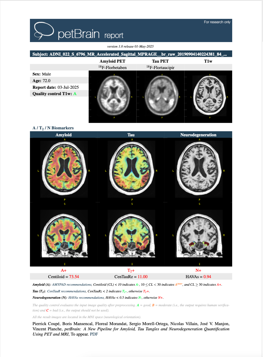
References
P. Coupé, B. Mansencal, F. Morandat, S. Morell-Ortega, J V. Manjon, N. Villain, V. Planche, petBrain: A New Pipeline for Amyloid, Tau Tangles and Neurodegeneration Quantification Using PET and MRI, Alzheimer's Research & Therapy (2025). PDF
P. Coupé, J. V. Manjón, B. Mansencal, T. Tourdias, G. Catheline, V. Planche. Hippocampal-amygdalo-ventricular atrophy score: Alzheimer disease detection using normative and pathological lifespan models. Human Brain Mapping 43, no. 10 (2022): 3270-3282. PDF
P. Coupé, B. Mansencal, M. Clément, R. Giraud, B. Denis de Senneville, V.-T Ta, V. Lepetit, J. V. Manjon. AssemblyNet: A large ensemble of CNNs for 3D Whole Brain MRI Segmentation. NeuroImage, 219, 117026, 2020. PDF
de Senneville, B.D., Manjon, J.V. and Coupé, P., 2020. RegQCNET: Deep quality control for image-to-template brain MRI affine registration. Physics in Medicine & Biology, 65(22), p.225022. PDF