CERES
CERES is a pipeline to automatically analyze cerebellum in brain MRI. It gets an anonymized T1w MRI brain volume in NIFTI format and produces a pdf report. CERES provides the volumes of the cerebellum lobules and the cerebellum tissues.
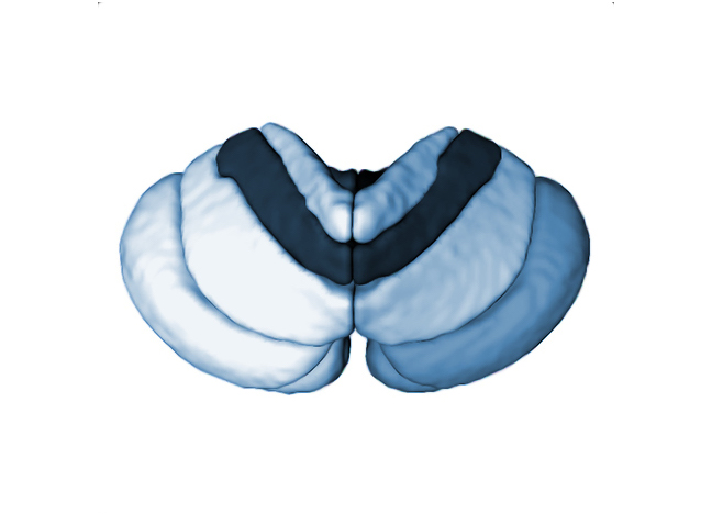
Labelling protocol
Cerebellum white matter and lobules were manually segmented by experts in a set of T1w MRI. This training library is then used to perform automatic segmentation of your new cases. All structures were outlined according to the definition described in Park et al., 2014.
Report
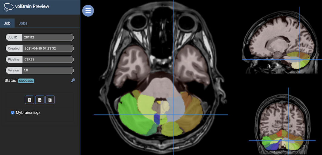
Once the process is finished, you will be notified by e-mail, so you will be able to download a package including some image files and two reports (CSV and PDF) providing all the volumes estimated from the segmentations. As you can see in the figure below, the PDF includes patient information, volume of cerebellum tissues and cerebellum lobules, cerebellum cortical thickness as well as asymmetry indexes. Finally, it also includes several snapshots from the different labeling steps as a quality control.
Download PDF Report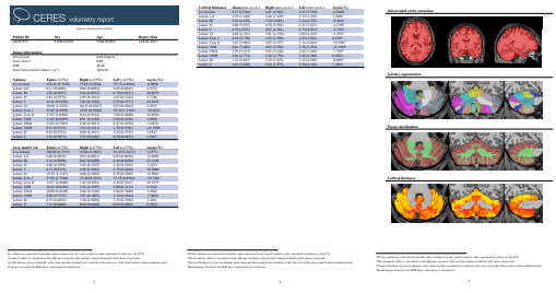
References
Jose E. Romero, Pierrick Coupé, Rémi Giraud, Vinh-Thong Ta, Vladimir Fonov, Min Tae M. Park, M. Mallar Chakravarty, Aristotle N. Voineskos, Jose V. Manjòn. CERES: A new cerebellum lobule segmentation method. Neuroimage, 147:916-924, 2017. PDF